자료는 저작권이 본인에게 있고 저작권에 문제가 없는 자료라면 판매가 가능합니다.
해피캠퍼스 메인 우측상단
[내계정]→[마이페이지]→[내자료]→[자료 개별등록] 버튼을 클릭해 주세요.
◎ 자료등록 순서는?
자료 등록 1단계 : 판매자 본인인증
자료 등록 2단계 : 서약서 및 저작권 규정 동의
서약서와 저작권 규정에 동의하신 후 일괄 혹은 개별 등록 버튼을 눌러 자료등록을 시작합니다.
①[일괄 등록] : 여러 개의 파일을 올리실 때 편리합니다.
②[개별 등록] : 1건의 자료를 올리실 때 이용하며, 직접 목차와 본문을 입력하실 때 유용합니다.
자료 등록 3단계 : 자료 상세 정보 입력
▶ 자료등록 가이드
1. 파일 선택 : [찾아보기]를 눌러 파일을 업로드합니다.
(등록가능한 파일형식 : doc, docx, ppt, pptx, xls, xlsx, hwp, hwpx, pdf, 압축파일)
2. 제목 : 자료의 내용을 파악할 수 있도록 구체적으로 기재하고, 불필요한 특수문자(★,☆ 등)는 지워주세요.
3. 카테고리 : 자료 유형에 맞게 선택해 주세요.
4. 과목명 : 리포트 과목명을 입력해 주시고 없으면 [과목명없음]으로 체크해주세요.
5. 판매가격 : 가격은 직접 책정해 주시고
무료자료는 추후 유료로 변경이 불가하니 주의해 주세요.
6. 저작시기: 해당 자료를 작성한 시기를 선택해주세요.
7. 태그 : 검색결과에 큰 영향을 줍니다. 따라서, 해당 자료의 검색에 도움이 되는 중요한 단어로 최대 10개까지 등록이 가능합니다.
8. 상세정보입력 - 검색 상위노출과 자료판매에 도움이 됩니다.
※일괄 등록하시거나 임의로 빈값으로 수정하는 경우 자료관리팀에서 정리합니다.
① 소개글 : 자료 구매 시 참고할만한 내용이나 특이사항이 있다면 자세히 기재해주세요. 자료에 대한 친절한 소개는 판매에 큰 도움이 됩니다.
② 목차와 본문 : 본문은 700 ~ 1,000자 내외로 입력합니다.
③ 참고자료 : 자료 내 기재되어 있는 참고문헌을 입력해주세요.
자료 등록 4단계 : 등록 완료
모든 정보를 입력하신 후 확인 버튼을 누르면 자료 업로드가 완료됩니다. 자료 업로드 완료 페이지에서는
현재
대기중인 자료 수와
검수 소요 예상 시간을 안내해 드립니다.
자료가 등록되면 자료관리팀에서 검수가 이루어지며
!등록 혹은 보류가 되면 회원님의 이메일이나 알림톡으로 해당 사실을 전달하여 드립니다.
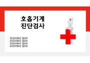
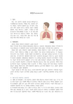
 호흡기계 진단검사, 호흡중재
4페이지
호흡기계 진단검사, 호흡중재
4페이지