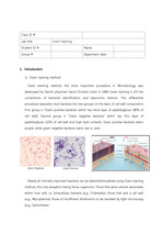
Gram positive bacteria stains purple, while gram negative bacteria stains red or pink. ... Class ID # Lab title Gram Staining Student ID # Name Group # Experiment date Introduction Gram staining ... Let stand for 60 seconds. Pour off the stain and gently wash with tap water from a faucet.
Immunofluorescence staining MATERIALS Chamber slide, PBS, 4% paraformaldehyde, 0.2% triton x-100, 1% ... for a few min or hour - Wash with PBS (3 times) - Incubate with 0.1-1 關g/ml Hoechst33342 or DAPI (DNA stain ... PROCEDURE - Count the cells and plating cells on each well of chamber slide (make single cell!)
H&E staining 외에 어떤 staining이 있고, 각기 어떤 용도로 쓰이는가. 2가지 대표적인 방법에 대해 설명하시오.1) Gram staining그람 양성균과 그람 음성균의 ... Eosin은 H&E staining에서 Hematoxylin의 대조 염색제로 주로 이용되며, Hematoxylin이 염색한 핵 등의 세포 소기관을 제외한 대부분의 구조를 붉은색으로
Gram staining도 H&E staining처럼 counter staining을 한다. ... H&E staining 외에 어떤 staining이 있고 각기 어떤 용도로 쓰이는가? 2가지 대표적인 방법에 대해 답하시오. ... 저배율에서 보았을 때, Sample 1 에서는 HE staining이 잘 되지 않은 하얀색 부분이 많이 보이지만, 청색으로 staining 된 핵이 선명하게 보이고, Eosin이 염색된
AP(Alkaline phosphatate) staining은 chemical의 반응을 이용한 staining이다. ... 일어나는 staining은 아니다. ... 그러나 실험 결과를 살펴보면 figure.2(a)da까지 staining이 되지 않아 AP staining 결과를 확인할 수 없었다는 추측을 할 수 있다.
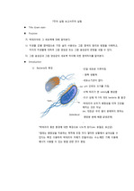
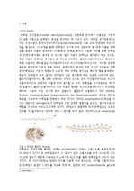
각각의 사진에서 공통으로 보이는 세로줄의 보라색 선은 spleen을 얇게 slice하는 과정에서 세포가 접혀 생겨난 현상이고, 사진1의 우측 상단에 보이는 마치 세포가 찢겨 나간 듯한 ... 사진4는 모든 처리 과정을 거치고 촬영하기 전에 찍은 spleen의 모습으로 보라색을 띄고 있다. 8. ... t cell과 plasma cell이 있다. red pulp에는 coros와 venous sinus가 있다.
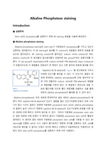
확인한다. ● Alkaline phosphatase staining Alkaline phosphatase staining은 stem cells가 미분화되어 pluripotency를 ... Alkaline Phosphatase staining Introduction ● 실험목적 Stem cell의 pluripotency를 검증하기 위해 AP staining 방법을 사용해 ... 이 AP staining의 원리를 각 solution의 성분들의 화학적 반응을 중심으로 알아보았다.
Causes of a hamstring strain Signs and symptoms of a hamstring strain Diagnosis of a hamstring strain ... hamstring strain What is a hamstring strain? ... They are commonly seen in running sports such as football, hockey and athletics (particularly sprinters
Therefore, this study was pursued to find out if Gram staining and fluorescent staining in addition to ... Therefore, if the Ziehl-Neelsen and Gram staining are combined as the M. tuberculosis staining method ... staining, culture, and nucleic acid amplification tests.
기본정보 실험 제목: 현미경 사용법 및 단순염색 (simple stain)과 그람염색 (gram stain) 실험 목적: 균의 형태, 크기, 배열상태 등을 관찰할 수 있다. ... 그람 염색의 과정과 결과 그람염색 법 / gram stain (tistory.com) 7. 제품실험자료 - (주)생물나라 (biozoa.co.kr) 8. ... (세포벽의 10~20% 차지) 그래서 크리스탈 바이올렛과 같은 염기성 염료로 염색한 후 에탄올을 처am stain) 1884년 덴마크의 미생물학자인 Hans Christain Gram이
Power supply를 꺼주고 tank를 꺼냅니다. #3. Gel 염색하기 Coomassie brilliant blue stain solution 1에 넣습니다. ... 후에 Coomassie Briliant Blue나 silver stain으로 염색한다. Marker 단백질을 사용하여 각 단백질들의 크기를 측정할 수 있다. *2. ... Coomassie brilliant blue stain solution을 회수합니다 증류수를 gel이 잠길만큼 넣고 5분동안 교반시킵니다.
단백질 염색은 대표적으로 본 실험에서 한 coomassie blue stain과 silver stain, ponceau S, specific antibody stin등이 있다. ... 그렇게 overnight한 후 staining solution에 gel을 20 min동안 넣어 staining시킨 후 염색약이 다 빠질 때까지 washing을 계속 진행한다. 3. ... 약 2시간정도 걸어준 후에 commassie blue staining을 진행한다.
단염색(simple stain), Gram stain, 항산성염색(Acid fast staining), 아포염색(Spore staining) 등이 그 종류이다. ... 본 실험에서 그람염색법(Gram staining)을 이용하였다. ... 우리가 이번 실험에 이용한 그람염색법 외에도 세균 세포를 단일 염료로 염색하여 세포의 형태와 배열을 관찰하고자하는 목적으로 사용되는 단염색(simple staining), 항산성을
의학 진단에서 널리 이용되며, 종종 최적 표준(gold standard)이다. ... - H & E stainingH&E 염색 혹은 HE 염색에서 H 는 헤마톡실린, E 는 에오신을 의미한다.1 조직학에서 이용되는 기본적인 염색 방법이다. ... 병리학자가 암으로 의심되는 조직을 생검하면 조직 절편을 H&E 염색하고, H&E 절편(H&E section)이라 한다.염색 기법은 헤마톡실린의 산화형인 헤마테인과 알루미늄 이온에서
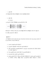
Transfer, Membrane staining / Cutting 실험 목적 : 전기영동을 통해 전개된 단백질들중 CYP2E1의 발현량을 알아본다. 실험 과정 #1. ... Transfer가 잘되었는지 확인하기 위해 membrane을 염색한다면 reversible staining방법을 사용해야 한다. : Coomassie brilliant blue : ... 그러므로 transfer에서는 stacking gel을 제거합니다) gel에서 투명한 실선이 stacking과 separating의 경계입니다. 이 실선을 잘라서 버립니다.